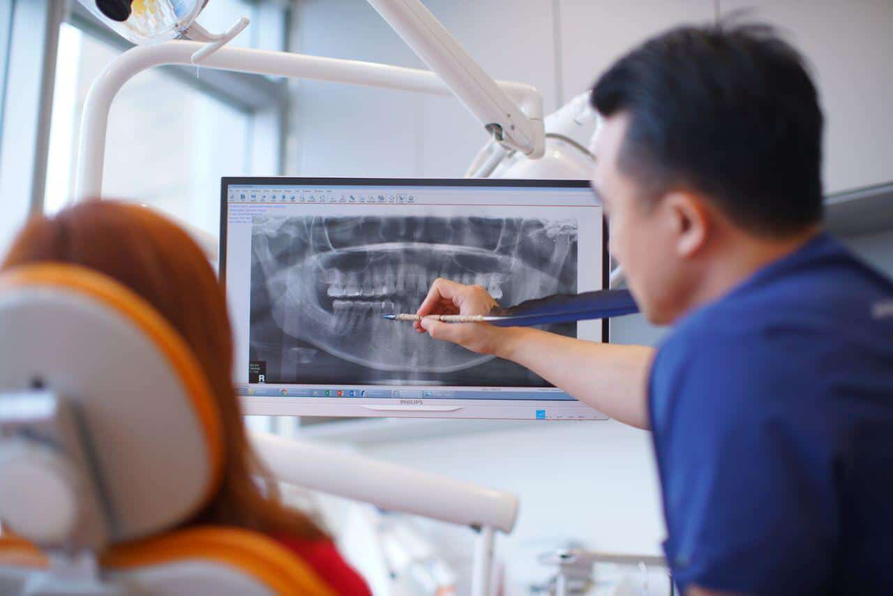
Digital dental radiology
X-ray illustration of teeth
Digital Dental Imaging
Elite Dental uses only digital radiology systems. The digital radiology images or x-ray images are “developed” instantly with a scanner.
Digital dental x-rays require only about 50% of the radiation dose used for traditional film x-rays. The radiation exposure is very minimal. We are aware that some of you may have the belief that dental x-rays are potentially harmful and can even cause an increased risk of cancers such as thyroid cancer.
We have created the table below to help dispel some of the myths regarding the perceived “ high radiation” dosages of dental x-rays.
| Procedure/source of exposure | Approximate radiation dose | Comparable to natural environmental radiation for: | *Additional lifetime risk of fatal cancer |
|---|---|---|---|
| CT Head Scan | 2 mSv | 8 months | Very Low |
| Mammography | 0.4 mSv | 7 weeks | Very Low |
| Chest X-ray | 0.1 mSv | 10 day | Negligible |
| Coast to coast flight in a commercial airplane | 0.03 mSv | ||
| 2 bitewing + 2 periapical dental x-rays | 0.008 mSv | 1 day | Negligible |
| Panoramic X-ray | 0.007 mSv | 1 day | Negligible |
According to the National Council on Radiation Protection and Measurements, the average person receives an effective dose of 3 millisieverts (mSv) per year from naturally occurring radioactive materials and cosmic radiation from outer space. Naturally occurring radioactive materials can be found in the air that we breathe in and the food that we eat. Cosmic radiation is that which we receive from the sun in the form of UVA/UVB wavelengths.
Why do I need to take dental X-rays?
X-rays are taken to detect underlying problems that cannot be seen just by looking into the mouth with the naked eye; for example, impacted wisdom teeth, bone loss due to gum disease, possible decay in between teeth and under old fillings, cysts and abscesses in the jaw etc.
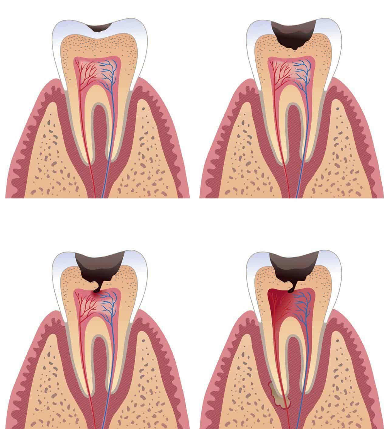
Illustration of tooth decay
Periapical X-rays (PA)
A PA x-ray takes a full tooth picture from the very top of the tooth (crown) to the very tip of the root and its surrounding bone. This is essential for detecting infections around the roots and is an essential x-ray for detecting root canal infections.
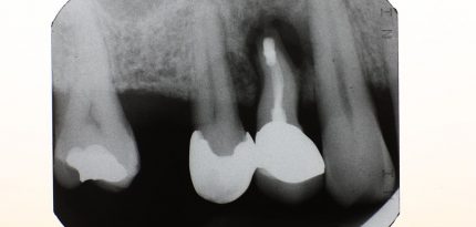
Periapical X-ray (PA)
Bitewing X-rays
Bitewing x-rays are taken to show the upper and lower molars (back teeth) and premolars (teeth in front of the molars). This is the best x-ray for detecting decay in between the back teeth.
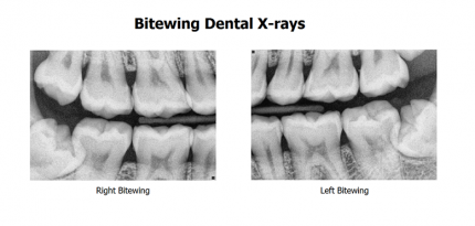
Left and Right Bitewing xrays
Orthopantomogram (OPG)
An OPG is a panoramic X-ray of the lower face, which shows all the teeth of the upper and lower jaws, as well as the surrounding bony structures (both maxillary sinuses, the nose, the cervical spine and the entire lower jaw and both jaw joints).
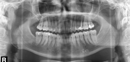
Orthopantomogram (OPG)
Lateral Cephalometric Radiograph (Lat Ceph)
A Lateral Ceph is a side view X-ray of the face, which shows the bones of the skull, neck and the teeth in profile. Lateral Ceph X-rays are typically used in the diagnosis of orthodontic problems.
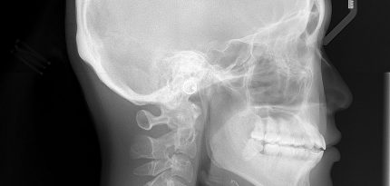
Lat Ceph
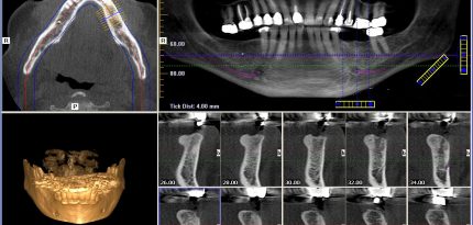
3D Cone Beam Computed Tomography (CBCT)
A 3D CBCT produces three dimensional (3-D) images of your teeth, maxillary sinuses, large nerves and bone in a single scan. Looking at these structures in 3-D can provide more spatial information beyond what a flat 2D x-ray would show, allowing us to plan in greater detail for more invasive and complex procedures. 3D CBCT scans are typically taken for wisdom teeth, dental implant and root canal related procedures.
Frequently
Asked
Questions
Got questions? We’ve got answers! Check out our FAQ section for common inquiries and helpful information to guide you.
What is digital dental imaging?
Digital dental imaging is a modern radiographic technique that captures dental X-ray images electronically rather than using traditional film. The two main technologies available are solid-state systems (using direct digital sensors) and photostimulable storage phosphor (PSP) plate systems.Digital radiographs are composed of numerical data arranged as a grid of rows and columns, which allows for computer-based image manipulation and enhancement.
Digital imaging has evolved from traditional two-dimensional (2D) imaging to advanced three-dimensional (3D) modalities such as cone-beam computed tomography (CBCT), overcoming many limitations of 2D imaging including superimposition and distortion.
What types of digital dental imaging are available?
The main types include intraoral digital radiography, digital panoramic imaging, and cone-beam computed tomography (CBCT). Intraoral digital radiography uses either direct digital sensors (most common, used in 68% of dental offices) or photostimulable storage phosphor technology (18%). Digital panoramic radiography is used in almost 80% of panoramic units. CBCT provides three-dimensional imaging of dental and facial structures with voxels as small as 0.125 mm.
Direct digital sensors offer immediate image production, while PSP plates require scanning after exposure but can be used for full-mouth series. Each technology has its strengths, and the choice depends on the type of dental practice and clinical needs.
What are the advantages of digital imaging over conventional film?
Digital imaging offers immediate image availability, reduced radiation exposure, enhanced diagnostic capabilities, and improved workflow. Key advantages include instant image production with solid-state devices, the ability to optimize brightness and contrast for specific diagnostic procedures, digital storage with easy access through practice management software, and the capacity to create perfect duplicates for referrals.
Image processing allows our team to enhance certain characteristics, correct overexposed or underexposed images, and perform advanced procedures such as digital subtraction radiography and computer-aided recognition of image features. Digital systems also eliminate the need for chemical processing and provide secure backup capabilities.
How much radiation exposure comes from digital dental X-rays?
Radiation exposure from digital dental imaging is very low and varies by examination type. For intraoral radiography, the effective dose is approximately 0.0050 mSv (5 μSv). Digital panoramic examinations deliver approximately 0.013 mSv (13 μSv), while CBCT scans range from 0.20 mSv (200 μSv) for standard protocols.
To put this in perspective, the effective dose from a single intraoral digital radiograph is equivalent to less than one day of natural background radiation. CBCT delivers higher doses than 2D imaging but remains much lower than medical CT scans. Thyroid doses from intraoral and panoramic imaging are minimal, with doses below detection levels for bite-wing and periapical radiographs.
How accurate is digital dental imaging for detecting cavities?
Digital imaging systems are as accurate as conventional film for caries detection, with sensitivities of 0.6 to 0.8 for detecting cavities into dentine. For approximal dentinal lesions, both sensitivities and specificities are fair, though detection of early enamel-only lesions remains challenging with all radiographic methods.
Overall, imaging for early caries detection shows a sensitivity of 0.47 and specificity of 0.88, meaning that while digital imaging is good at confirming the absence of disease, it may miss some early lesions. Digital systems perform as accurately as film-based imaging despite having more limited resolution, and the ability to enhance images may improve diagnostic accuracy.
HOW CAN WE
HELP?
Heading to a dental clinic can be a scary and almost overwhelming experience. If you have landed on our website, we would like to extend our warmest welcome to you and your family. There is nothing to fear!
If you require more information about our costs or are looking for ways to save money on dental treatments, arrange for a consultation with our dentists at a time of your convenience.